Don’t follow the dollars, follow the data
The future of surgery should not and will not be written by technologists alone. It must be co-authored by us surgeons. To all my colleagues, I would say, don’t just be a user of the future; be an architect of it.’
These were the words of Dr Zubin Daruwalla, a former orthopaedic surgeon whose career path has been an interesting one. After training in his speciality, he left full-time clinical practice, frustrated with the system, to join PricewaterhouseCoopers (PwC) to oversee the Asia Pacific Health Industries practice and the global health leadership team. He now enjoys what he calls a hybrid career.
A doctorpreneur, surgeon and futurist, he was among the guest speakers who shared insights with delegates recently at The Surgeon Show.
With extensive experience across clinical practice, digital health, and innovation, he reflected on his experience spanning Asia and the US, examining how diverse health systems, cultures, and expectations shape clinical practice and professional development.
Speaking with the Show’s media partner, Future Medicine, following his well-received session, Zubin said that bringing together surgeons was ‘extremely valuable’.
‘We share the same mindset. We get to see what is happening in the surgical field, so I think bringing the community together has been absolutely fantastic. I am loving hearing about the cultural experiences of people from around the world, and about what different systems are doing, because we can learn from them.’
His buzzword for surgeons was ’glocalise’: learn globally, then adapt locally.
A decade ago, with shifting career goalposts, Zubin felt frustrated. Returning to Singapore after practising in the UK was challenging; the country’s postgraduate medical education, including surgical training, had been overhauled, replacing the British-style system and requiring him to retrain.
Yet, his primary motivation was to have a bigger impact on just the patient in front of him; he wanted to affect populations and healthcare systems.
At PwC, he helped hospitals in Asia with electronic medical records, amalgamated entities into an academic medical centre, worked on financing, and also set up biotech cities in the Middle East and residency programmes worldwide.
It offered him breadth and depth – not just one patient at a time.
Consequently, he suggested that if you silo yourself solely as a surgeon, you may miss out on additional opportunities to do more.
In medicine, specifically in surgery, you don’t realise that to become good at what you do, you go into a speciality and do more and more of the same thing, but the problem is you become so narrow you cannot do anything else, he observed.
Surgery, he felt, does not allow for variety. Now, younger generations want more time for themselves and their families; they want to travel and work fewer hours.
Zubin said: ‘The onus is on us. We need to keep up with the demand. We have to evolve, and we have to adapt.’
One caveat is that, in healthcare, it’s a profession where the more you do, the better you get, so it’s a difficult balance to strike.
He asked: ‘How do we balance spending enough to gain the right experience to become competent in delivering care, while meeting these new expectations?’
A hybrid career seems to be the answer.
So, how does medical technology enhance and augment that?
‘Looking at it from the perspective of the patient journey, in any specialty, we start with awareness and education for the individual and the population about the disease, then move to prophylactics, e.g., vaccinations, then diagnostics, then therapeutics, then follow-up care,’ he explained.
‘Dividing up technologies, we look at software solutions, software as a medical device, hardware solutions, and then molecules (pharma, etc.). This forms a matrix in which technology cuts across the entire delivery of care.’
One final note of caution, ‘don’t get caught up with the hype,’ he added. ‘Don’t follow the dollars, follow the data. Be cautious in your approach,’ he warned.
With an impressive attendance of 656 C-suite surgeons from around the world, the UK’s pioneering Surgical Leaders Summit has cemented its place on the global surgical calendar.
Looking ahead, The Surgeon Show 2027 UK’s Exclusive Surgical Leaders Summit will take place at London’s Business Design Centre on Friday 19 January.
The Surgeon Show highlights an ‘invisible problem’ in the OR
Medtech has long overlooked the ‘invisible problem’ of gender health and data gaps. Device development must break from traditional norms if companies are to promote inclusivity.
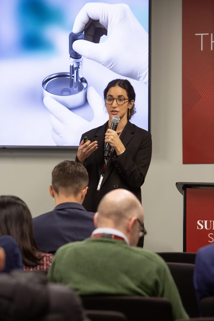
Lara Zaki, senior FEI strategy and innovation consultant at Team Consulting, shared this view at The Surgeon Show, a pioneering event attended by over 656 leading surgeons worldwide to discuss innovation, change, and the future of surgery.
A highlight of the exclusive Surgical Leaders Summit was hearing from innovators transforming the surgical environment.
Team Consulting is an award-winning medical device consultancy with 40 years of experience in medical product design and development. It actively raises awareness of these gaps by employing cognitive science in early-stage medical device strategy, leads femtech initiatives to reduce gender health disparities, and supports women’s health innovators.
Lara Zaki co-presented a session with Shweta Aggarwal, a consultant plastic surgeon at Bart’s Health and an Honorary Senior Lecturer at QMUL, in which they discussed Team Consulting’s work to help develop a novel device concept to improve surgeon accuracy in breast surgery procedures.
The talk reflected another key theme throughout The Surgeon Show: that surgeon-led innovation is critical for effective solutions, relying on collaboration among clinicians, industry, and surgical bodies.
Following the session, Lara spoke with the Show’s media partner Future Medicine and explained that women surgeons frequently face problems in the operating room and are often told to ‘just figure it out’ when they encounter issues with instruments.
She shared that she’s heard many stories highlighting how devices are not suitable for smaller hands, varying hand strengths, or different heights, affecting both male and female surgeons. This, she added, is a genuine health concern that impacts a surgeon’s wellbeing and their ability to perform efficiently and effectively.
Team Consulting aims to close these data gaps and ensure that device development genuinely includes all users.
However, developing data is often hindered by limited access to information that could make products more inclusive and better address women’s health issues in a male-dominated industry. This gender bias persists not only in device development but also in how students are taught to use them.
Lara also noted a tendency to rely on precedent in medical device development. Many devices and medical textbooks are based on male anatomy, perpetuating this ongoing gap, she said.
Recently, Team Consulting conducted an interactive workshop to explore and address these issues of inclusivity. The session, held at the Women at the Cutting Edge event, hosted by the Royal College of Surgeons of England in partnership with the RCSEng’s I-Hub and Women in Surgery (WinS), brought together surgeons to tackle gender bias in surgical device design.
Co-founder of The Surgeon Show, Professor Shafi Ahmed, said: ‘Meaningful dialogue drives progress. Together, we are shaping the next era of surgery, with innovation, responsibility, and patients at the centre of every conversation. Let us think boldly, lead responsibly, and build what comes next together.’
‘You can go from being a resident one day to being a consultant the next day. What extra skills have they taught you in those 24 hours? Probably not a lot.’
This was the insight shared by Professor Fiona Myint recently, urging the profession to incorporate surgical leadership skills at all levels.
Speaking at The Surgeon Show in London, she participated in a panel exploring how leadership demands are evolving with advancements in surgery.
And she argued that the surgical profession ‘will thrive so much more if we are at the helm’.
The exclusive Surgical Leaders Summit brought together leading surgeons from around the world to discuss innovation and its impact on the profession.
The event highlighted an urgent need for leadership to adapt amidst rapid innovation, drawing 656 C-suite surgeons from across the globe.
‘Leadership in Contemporary Surgical Practice’ examined what is required to lead surgical services at scale, balancing workforce, quality, innovation, performance, and accountability in an increasingly complex healthcare environment.
Chaired by Professor Domenico Veneziano, the panel included Professor Fiona Myint, clinical professor of surgical education at Royal Free Hospital, Peter Gogalniceanu from Guy’s & St Thomas’ NHS Trust, and Professor Nora Colton, director at ULC Global Business School.
From managing high-stakes teams to navigating structural change and setting the tone for excellence, the discussion focused on practical lessons in influence, resilience and strategic thinking.
Speaking with the event’s media partners, Future Medicine, Fiona Myint emphasised the importance of understanding what it is like to be a leader, especially in the context of innovation.
She said: ‘We don’t teach leadership, especially lower down the echelons, and we must incorporate it. I set up the Harvard surgical leadership course to teach consultation surgeons about leadership, because it isn’t discussed in training. I strongly believe we should do this at the trainee and medical student levels because, as you climb the surgical ladder, while you learn to operate and treat patients, when you are thrown into a junior leadership role, you’ve never been taught how to do it. You don’t know how to read spreadsheets, understand budgets, or lead a team, and these skills need to be taught earlier. As a profession, if we take on the leadership roles, then we’re leading our own profession. Our profession will thrive so much more if we are at the helm.’
Change is happening around leadership, she admitted, but not necessarily in a structured way, so this event also served as a call to action.
‘You can go from being a resident one day to being a consultant the next day. What extra skills have they taught you in those 24 hours? Probably not a lot,’ she added.
She listed emotional intelligence, situational awareness, and knowing who it is you’re leading and what you are leading for as core values. We need to be there, leading the team, leading the institution to give the best possible care to our patients, she said. Patient gets the right care from a happy team.
She praised the choice of keynote speaker, Rafael J Grossmann, the widely acclaimed general and trauma surgeon and pioneering digital health innovator, who, she said, ‘opened everybody’s eyes and enthused everybody from the start’.
Feedback was overwhelmingly positive. Among the delegates was Ginny Bowbrick, consultant vascular surgeon and head of the Kent, Surrey & Sussex School of Surgery, who said: ‘A fabulous event bringing together our community to discuss advancing innovation in surgery across a wide spectrum, including, most importantly, workforce and training. We need to grasp the opportunities and challenges now, or we will be left behind in the global arena.’
Dr Rafael J Grossman leads the discussion at The Surgeon Show
‘We’ve been displaced by technology in healthcare, and we are forgetting about the human patient in front of us.’ That was the stark warning from Dr Rafael J Grossmann, renowned trauma surgeon and digital health pioneer, who was the keynote speaker at The Surgeon Show in London last month.
Celebrated for his innovative approach at the crossroads of healthcare and technology, Dr Grossmann captivated the audience with his passionate speech, emphasising the event’s importance as a pioneering platform for surgical innovation and collaboration.
He praised the event as ‘magnificent and most-needed’, expressing enthusiasm for how it showcased new robotic technology and AI applications in healthcare, as well as its facilitation of the ‘meeting of global minds’.
However, his main message was a cautionary one: implementing new technology in surgery cannot happen without considering the human element.
Dr Grossmann, a self-confessed ‘geek,’ was one of the earliest adopters of wearable technology in the operating room and has long been an advocate for digital innovation in surgery. He said he has ‘always been fascinated by the power and potential of technology to change how we do things’.
But he warned that ‘technology pushes us to forget the person in front of us’. For him, using AI, robotics, and new technologies intelligently means holding on to our humanity.
Speaking with the event’s media partners, Future Medicine, after his address, he said it was important to ‘use technology smartly in order to not forget that the primordial goal is that person in front of you’.
‘Technology is almost forcing us to do that unless we fight back,’ he added. ‘We forget to talk, we forget to inform. And the patient becomes a number, becomes a case, becomes a disease pathology rather than a human with disease.’
He also suggested that the profession itself can sometimes act as a barrier to the adoption of new technology. ‘There are cost barriers, there are regulatory barriers, and there are political barriers. But education is the main barrier – for the professionals, the regulators and the administrators. I’m a full-time evangelist, and if we don’t make it our goal to really change minds, it will never happen… we need to accelerate it because we have no time.’
He agreed that there can be a disconnect between surgical training and what medical students are exposed to, as well as the realities of clinical practice, and emphasised that the profession needs to update the curricula. The way surgery is taught, even in countries in the Global North, is decades out of date, he said, adding that it is a ‘paradigm that we need to figure out how to change’.
‘In the five to seven years of training, the world changes radically. Every day, there is something new in AI and robotics, so unless this is adapted and brought to the training of a new generation, playing catch-up only proves difficult and costly, contributing to burnout and affecting patient care.’
Elsewhere, the day’s discussions spanned all aspects of future surgery, including robotics, AI, and immersive technologies, as well as digital ethics, data-driven care, training, leadership, and mentorship in the modern era.
Attendees could access 25 curated, high-level sessions on recent advancements and challenges in surgery, offering excellent networking opportunities.
The exhibition, complementing the sessions, showcased a wide range of innovative surgical technologies, including artificial intelligence, digitalisation, virtual reality, 3D printing and robotics.
Successfully attracting an impressive 656 C-suite surgeons from around the world, the UK’s pioneering Surgical Leaders Summit has already cemented its place on the global surgical calendar.
‘The Surgeon Show truly sets the tone for what lies ahead, showcasing the very best of global robotic surgical practice with a clear focus on improving patient outcome’
London hosted the first-ever Surgeon Show on 20 February, igniting innovation, sparking collaboration and transforming the surgical community, earning itself a place on the global surgical calendar.
According to one guest, the pioneering and exclusive Surgical Leaders Summit ‘sets the tone for what lies ahead, showcasing the very best of global robotic surgical practice with a clear focus on improving patient outcomes’.
Designed to shape the future of surgery, drive change and elevate leadership within the profession, The Surgeon Show 2026 was held at the prestigious Minster Building in the heart of London’s financial district and attracted an impressive 656 C-suite surgeons from around the world.
It featured more than 60 expert speakers and distinguished faculty, drew clinicians from across the globe, and was supported by over 50 healthcare organisations.
The event provided an exceptional platform for learning, sharing and collaboration and, thanks to its impressive line-up of speakers and guests, it was perfectly placed to disseminate insights from some of the most influential voices in the sector.
Attendees participated in 25 curated, high-level sessions that addressed the latest advancements and challenges in surgery, offering exceptional networking opportunities.
The complementary exhibition showcased a wealth of cutting-edge surgical innovations, including those in artificial intelligence, digitalisation, virtual reality, 3D printing and robotics.
The keynote speaker was Rafael J Grossmann, the widely acclaimed general and trauma surgeon and pioneering digital health innovator, whose passion for the intersection of healthcare and technology was palpable in his address.
Hailed as a top influencer in digital health transformation, he described The Surgeon Show as a ‘magnificent and most-needed event’.
Among the key themes covered, delegates were treated to sessions on:
• The future operating theatre – integrating robotics
• AI and immersive technologies
• Digital ethics and data-driven care in an AI-enabled world
• Training, leadership and mentorship in the modern era.
Speaking after the event, Professor Shafi Ahmed, the multi-award-winning surgeon, educator, and innovator, and co-founder of Surgery International and The Surgeon Show, admitted he was overwhelmed by the response.
‘Thank you to all who came and supported The Surgical Leaders Summit at The Surgeon Show, which has found its permanent place in the surgical calendar,’ he said. ‘When we set out to build an entirely new event, we looked at the landscape and knew it was necessary. Our sole purpose was to connect senior surgeon leaders with industry partners and policymakers. We succeeded in our endeavours by creating an inspiring environment for the ultimate decision-makers to meet in one room to rethink, reimagine and redesign the future of our profession.’
He added: ‘Surgery presents unique challenges, and we wanted to take our guests on a journey that would push them out of their comfort zones and expand their minds and horizons – to create a different way of thinking and bring global perspectives. From mind-blowing keynotes to insightful panel discussions, from robots and exoskeletons to extended reality and AI surgical platforms, the agenda was created to inspire.’
Zameer Shah, a consultant orthopaedic surgeon at Guy’s and St. Thomas’ NHS Foundation Trust, summarised the mood: ‘Massive credit goes to Professor Shafi Ahmed and Mr Tim Lane (RCS President Elect), and the entire team who organised this visionary, informative, and collegial gathering, bridging the interfaces of surgery, healthcare, innovation and technology.’
He added: ‘From the powerful keynote by Dr Rafael J. Grossmann to the concluding reflections by Professor Aymeric Lim, Singapore, this was a truly international and inspiring event. It was a privilege to attend.’
Indeed, the feedback was overwhelmingly positive. Ryan Kerstein, surgeon and NHS innovation leader, remarked: ‘Thanks for creating The Surgeon Show as a space for honest conversations. Congratulations on delivering something genuinely different.’
Nikhil Vasdev, professor and chair of robotic surgery (HMS, UH), added: ‘Thank you for organising such a remarkable academic surgical event. The Surgeon Show truly sets the tone for what lies ahead, showcasing the very best of global robotic surgical practice with a clear focus on improving patient outcomes.’
Tim Lane, consultant urologist and robotic surgeon and the Royal College of Surgeons’ president elect and editor in chief of Surgery International, who co-founded the event, said: ‘Thanks go to all the global sponsors, exhibitors, and speakers, whose insights and expertise are shaping the future of surgery, who share our ambitions in helping build our responsible future together. Ultimately, we wanted to create something new, unique, disruptive and special.’
Professor Shafi added: ‘In the world of social media, visibility and brand awareness, we wanted to create lasting and powerful media, and our media team partnered with the brilliant team from Future Medicine. Together, we are shaping the next era of surgery, with innovation, responsibility and patients at the heart of every conversation. Let us think boldly, lead responsibly and build what comes next, together.’
Other sponsors were thanked, too, for supporting Surgery International in bringing The Surgeon Show to life. They included:
• Albright IP
• AV Medical Systems
• Cambridge Consultants
• CMR Surgical
• Concentric
• The Confederation of British Surgery
• DarwinGroup
• Erbe
• Healthcare Skills Training International
• J&J MedTech
• Medtronic
• Module CO
• Orascoptic
• Organa
• R2 Surgical
• Regency Wealth Management
• The Royal Society of Medicine
• Stanley
• Team
• VitVio.
An exoskeleton designed to reduce strain and combat muscular fatigue during long hours in the operating room could ease surgeons’ stress.
Kapil Sahnan, a UK colorectal surgeon at St Mark’s The National Bowel Hospital in London, is the first to wear the device during a seven-hour procedure.
The exoskeleton suit is worn over scrubs and designed to support the arms and shoulders during prolonged surgical procedures.
He said: ‘Surgery can be physically demanding, especially during longer procedures, which can last many hours and being hunched over an operating table is not great for your posture. This is a welcome piece of kit for surgeons.’
Andre Jutel from Stanley, specialist experts in this area, assessed Kapil’s movement using their ErgoScan app before the procedure.
The Hapo Front exoskeleton was recommended because it helps reduce fatigue and supports surgeons in working comfortably and with precision.
The lightweight exoskeleton, which comprises adjustable back, shoulder and arm straps, supports the wearer's musculoskeletal structure and maintains freedom of movement without restriction.
Kapil said: ‘It’s good because you can maintain the dexterity of your hands while performing surgery. During the training, I did feel like I was being measured for a suit, but the exoskeleton is surprisingly easy to put on and adjust. It is certainly something I will be using again. We’re a specialised hospital with a small niche workforce, so we can’t afford to have surgeons going off sick with issues like muscular disorders, which is a common side effect of the profession.’
Graham Sharp, Managing Director of Stanley, added: ‘The fantastic feedback from Mr Sahnan and his team demonstrates how exoskeleton technology is supporting the profession, particularly during lengthy operations. We advised the team at St Mark's on the best technology for this type of procedure and supplied training along with the HAPO Front exoskeleton.
‘AI-powered wearable technology is already revolutionising professional workflows across the healthcare sector, significantly mitigating physical fatigue. We are moving toward a future where exoskeleton equipment will be a standard fixture in operating theatres throughout the UK.’
A gathering of some of the most respected and forward-thinking minds in surgery will take place at The Surgeon Show 2026 on Friday, 20 February, at The Minster Building, Central London.
Positioned as one of the UK’s most influential surgical gatherings, this invitation-only, one-day summit will convene consultant surgeons, clinical directors, policymakers, healthcare innovators and technology leaders from across the UK and around the world.
Together, they will explore how robotics, artificial intelligence and advanced technologies are redefining modern surgical practice and shaping the future of patient care.
Headline partner Darwin Group supports the event, bringing its expertise in designing and building permanent-grade, on-demand healthcare facilities that enable trusts and organisations to deliver exceptional care.
The Surgeon Show 2026 has been awarded Ethical MedTech Accreditation and international CPD (Continuing Professional Development) accreditation by The CPD Certification Service. The accreditation recognises the event’s educational quality and provides formal professional recognition for attending surgeons worldwide.
Accredited delegates will receive five CPD points, contributing towards professional development requirements for regulatory bodies and membership organisations in the UK and internationally.
Designed as a curated, high-impact forum, the summit will bring together frontline clinical insight, system-level strategy and real-world innovation at scale. The programme reflects the rapid pace of transformation across surgery and healthcare systems worldwide.
Event highlights include:
- 60+ expert speakers and faculty
- 50+ healthcare organisations represented
- 25+ curated high-level sessions
- Three live theatres running concurrently
Professor Shafi Ahmed, multi-award-winning surgeon, educator and global innovator, said:
“The Surgeon Show is more than an event, it’s a movement. We’re creating a space where the brightest minds in surgery can come together, challenge each other and shape the future of our profession. Every voice here matters, because the future of surgery depends on collaboration across disciplines, generations and technologies.
We are delighted to welcome more than 60 expert speakers from across the UK and internationally, representing over 50 NHS trusts, private providers, universities and global health systems. Across more than 25 curated sessions, with faculty from Europe, North America, Asia and the Middle East, the programme reflects the breadth and pace of change in surgery today.”
Among the standout sessions are:
- Scaling Robotic Surgery Across International Health Systems, chaired by Professor Bijen Patel, exploring global implementation and sustainable adoption of robotic programmes.
- Controlling Incisions: An Agile Device Innovation Journey, with Team Consulting, examining how clinical need is translated into viable surgical devices through interdisciplinary innovation.
- The NHS 10-Year Plan Through the Lens of Robotic Surgery, chaired by Ms Niyati Lobo, exploring how policy, workforce planning and investment will shape robotic surgery over the next decade.
- AI-Guided Surgery: Decision Support, focusing on how AI-driven tools are entering the operating theatre alongside governance, safety and evidence considerations.
- Innovation Spotlights, highlighting emerging technologies including AI-powered operating rooms, surgical intelligence platforms, digital consent, advanced imaging and extended reality.
Unlike traditional conferences, The Surgeon Show is designed to be conversational and collaborative, encouraging cross-disciplinary dialogue and practical insight into how technology and teamwork are transforming surgical practice.
Key themes for 2026 include:
- The future operating theatre - integrating robotics, AI and immersive technologies
- Digital ethics and data-driven care in an AI-enabled world
- Training, leadership and mentorship in the modern era
Created to unite and inspire the surgical community, The Surgeon Show is a by-invitation-only summit for leaders shaping the next era of surgical care.
Confirm your complimentary place here:
Medtronic will bring its next-generation surgical robotics offering to The Surgeon Show, where attendees can see live demonstrations of the Hugo™ Robotic-Assisted Surgery (RAS) system alongside the company’s Touch Surgery™ digital training and education platform.
The appearance comes as Medtronic continues to expand the footprint of Hugo™ RAS internationally. The modular and portable system, developed for soft-tissue surgery—is now FDA cleared in the United States for urologic procedures and CE-marked for soft tissue abdominal procedures including urological, gynaecological, colorectal and general surgeries in Europe, positioning it as an increasingly prominent option in the fast-moving global robotic surgery landscape.
At the centre of Hugo™ RAS is a modular design built around independent arm carts, enabling teams to configure the system from one to four robotic arms depending on the needs of each case. The platform also features an open surgeon console, intended to support greater communication and coordination between the surgeon and the wider operating theatre team.
Medtronic says the flexible set-up supports multi-quadrant access, allowing teams to tackle more complex procedures while helping maintain smooth workflow - particularly in environments where space, set-up time and adaptability are key operational priorities.
Vanessa Lowe, Senior Business Unit Director, Medtronic Surgical, UK and Ireland: “At The Surgeon Show, we’re looking forward to demonstrating how Hugo™ RAS has been designed to fit into real clinical environments, bringing modularity, flexibility and choice to robotic-assisted surgery. By integrating Touch Surgery™ training and post-operative analytics, we’re also helping teams build skills and insight that support continuous improvement, both in and beyond the operating theatre.”
Alongside the robotics hardware, Medtronic will also highlight integration with Touch Surgery™ Enterprise, a cloud-based platform that combines interactive training content, surgical video recording, live streaming, and post-operative analytics designed to support surgeon education and performance improvement.
The company will also showcase the LigaSure™ Maryland RAS vessel-sealing instrument, CE-marked in 2025 for use with Hugo™ RAS, bringing its established vessel‑sealing capability into a robotic‑assisted setting.
Visitors to Medtronic’s booth at The Surgeon Show will be able to see the Hugo™ RAS system in action, alongside demonstrations of Touch Surgery™ live streaming, providing a hands-on view of how robotic platforms, training tools and analytics are increasingly converging to support modern surgical practice.
The Surgeon Show 2026 will bring together senior surgical leaders and industry innovators for a programme shaped around conversation and debate, exploring the issues influencing the future of surgery - from the operating theatre of tomorrow to the growing role of AI, robotics and advanced technologies in clinical practice.
The event is invitation-only, with registration of interest available via surgeonshow.com.
Disclaimers
The Medtronic Hugo™ RAS system is commercially available in certain geographies. Regulatory requirements of individual countries and regions will determine approval, or market availability. Indications for use may vary. Risks may include arrythmia, bleeding, burns, infection, tissue damage or other complications.
The Medtronic Touch Surgery™ ecosystem is not intended to direct surgery, or aid in diagnosis or treatment of a disease or condition. Please note: Performance Insights are available for select procedures, instruments, and anatomy.
The Surgeon Show 2026, with Healthcare Skills Training International, has been awarded international CPD (Continuing Professional Development) accreditation by The CPD Certification Service.
The accreditation confirms the event’s educational quality and provides formal professional recognition for attending surgeons worldwide.
The one-day conference takes place on Friday, 20 February 2026, at The Minster Building, London, bringing together surgeons, healthcare innovators and technology leaders to explore the future of surgical practice.
Accredited delegates will receive five CPD points, which can be used towards CPD requirements for professional bodies, regulators and membership organisations in the UK and internationally.
Professor Shafi Ahmed, one of the hosts of The Surgeon Show, said: “Securing CPD accreditation with Healthcare Skills Training International (HSTI) validates the rigour of our programme and ensures surgeons receive meaningful recognition for their professional development.”
The Surgeon Show 2026 programme focuses on the clinical, technological and leadership challenges shaping modern surgery, including:
- Robotic surgery and the future operating theatre
- Artificial intelligence in surgical practice
- Immersive technologies such as VR, holography and avatars
- Leadership and innovation in healthcare systems
The programme features internationally recognised speakers including Professor Shafi Ahmed, Professor Nikhil Vasdev, Professor Aymeric Lim and Professor Yujia Gao, alongside senior leaders from NHS trusts and global medtech companies.
The event will run across three concurrent theatres and include keynote presentations, panel discussions, innovation spotlights, industry-led sessions and structured networking opportunities, including breakfast sessions and an evening reception.
To achieve CPD accreditation, HSTI demonstrated that The Surgeon Show 2026 meets strict quality assurance criteria, including:
- Delivery of structured, high-quality educational content
- Appropriately qualified speakers and faculty
- Clear learning objectives and measurable outcomes
- Professional standards aligned with healthcare sector requirements
- Tangible professional development value for attendees
Tim Lane added: “This accreditation reflects the commitment of our advisory board and speakers to advancing surgical knowledge and practice. Our ambition is to establish The Surgeon Show as the premier CPD-accredited event for the global surgical community.”
Healthcare Skills Training International is dedicated to providing professional clinical education to post-graduate clinicians, allied healthcare professionals and device industry product support professionals. We believe that everyone in an acute care setting should have formal training to support patient safety, all ethical and legal requirements and clinical effectiveness.
All registered delegates will receive a CPD Certificate of Attendance after the event.
The Surgeon Show 2026 will bring together senior surgical leaders and industry innovators for a programme shaped around conversation and debate, exploring the issues influencing the future of surgery - from the operating theatre of tomorrow to the growing role of AI, robotics and advanced technologies in clinical practice.
The event is invitation-only, with registration of interest available via surgeonshow.com.
Cambridge Consultants, the deep tech powerhouse of Capgemini, has been confirmed as sponsor of the opening breakfast session at The Surgeon Show 2026, bringing together surgeon-innovators to discuss what it takes to turn clinical insight into scalable medtech solutions.
The session, titled “The Key to MedTech Success with Cambridge Consultants”, will take place at 8:30am and will examine the innovation-to-commercialisation journey from identifying an unmet need in surgery, through product build and investment, to revenue generation, adoption and growth.
Chaired by Oliver Sowerby, Vice President of MedTech Innovation at Cambridge Consultants, the breakfast panel will feature clinical innovators at different stages of company and product development, including:
- Mr Ali Haddad, CEO and Founder, XRlabs
- Ms Emily Mills, CEO and Co-founder, Cascade MedTech
- Mr Ash Kalraiya, CEO and Founder, Medishout
- Ms Terouz Pasha, Inaugural Innovation Fellow, London Institute of Healthcare Engineering & RCS Innovation Hub
- Mr Alex Almoudaris, CEO and Founder, TouchPoints.Health
The session will open with a short talk, followed by a panel discussion focused on a core theme: why clinical workflow matters and why even well-funded projects can fail if technologies don’t work in real surgical settings.
Commenting on the session, Oliver Sowerby said: “Surgeon-innovators are uniquely positioned to identify unmet needs, but building a company around that insight brings new challenges from engineering and regulation to investment and adoption. We’re proud to support this session and help create an open, honest conversation about what success really looks like in medtech.”
The panel will explore how surgeons are translating frontline insight into technology-led ventures, and the practical challenges that emerge as innovations scale — including product development, regulatory pathways, funding, clinical adoption and commercial growth.
The Surgeon Show 2026 will bring together senior surgical voices and industry leaders for a programme designed around conversation and debate, covering topics shaping the future of surgery - from the operating theatre of tomorrow to the growing role of AI, robotics and advanced technologies in clinical practice.
The event is invitation-only, with registration of interest available via surgeonshow.com.